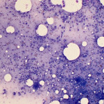
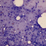
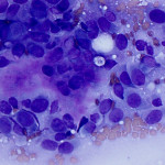
Aspirate from the distal femur of a dog
Case information
An 8 year-old, female spayed Rottweiler was presented to the Cornell University Hospital for Animals Emergency Service for a 6 day history of left hind limb pain. The dog was initially presented to the referring veterinarian 3 day prior for acute lameness and pain, which were unresponsive with Tramadol and Rimadyl treatment.
The dog was bight and alert and physical examination was unremarkable except for the left hind limb lameness. On sedation, there was decreased maximum flexion of the left elbow with crepitus, decreased range of motion of the left hindlimb, decreased maximum extension of the hips bilaterally, and effusion of both stifles, which was more prominent on the left stifle. A firm, painful swelling was identified on the medial and lateral aspects of the left hind limb, just above the stifle. Radiographs of the left hindlimb was performed and revealed an aggressive bone lesion on the distal femur with some proliferation. A CBC and chemistry panel were unremarkable except a marginally increased amylase activity (1259 U/L; reference interval: 337-1220 U/L) and mildly increased cholesterol concentration (360 mg/dL; reference interval: 138-332 mg/dL).
A fine needle aspirate was taken from the lesion on the left distal femur and submitted to Clinical Pathology.
- Based on the clinical presentation, what would be your top differential diagnosis?
- How do you classify the cell population in Figures 1 to 3?
- What is the nature of the extracellular material in Fig 3?
 |
 |
 |
Answer on next page
