
A left shift indicates the presence of immature neutrophils in blood and usually, but not always, indicates an inflammatory leukogram (see related links for the historical origin of this term). Immature neutrophils are usually band neutrophils, but earlier forms can be seen. A few to no band neutrophils are seen in the blood of clinically healthy animals we use for establishing our reference intervals. This indicates that low numbers of band neutrophils, particularly in the absence of other features of inflammation, such as toxic change, may not be a clinically relevant finding. Some concepts associated with a left shift:
- A left shift can be due to release of bone marrow stores. This particularly occurs if the bone marrow reserve of mature neutrophils is low or depleted. Note, that ruminants have lower bone marrow reserve of mature neurophils than other species. Release of the marrow reserve usually occurs in response to acute inflammation. Stores will be rapidly depleted if bone marrow production (resulting in a myeloid or granulocytic hyperplasia) does not kick in – this marrow response usually takes around 3-4 days and occurs due to the release of granulopoietic cytokines, such as granulocyte- colony stimulating factor (G-CSF), granulocyte-monocyte-colony stimulating factor (GM-CSF) or inflammatory cytokines such as interleukin (IL)-1 and IL-6.
- A left shift can indicate a response by the bone marrow to inflammatory or granulopoietic cytokines, which increase marrow production, i.e. there is a myeloid (granulocytic) hyperplasia.
- In a single hemogram, we cannot always tell if there is a myeloid hyperplasia. A general rule of thumb is that if there is a persistent left shift with a neutrophilia, there is likely a myeloid hyperplasia. If the neutrophil count is very high (>40,000/uL in a dog or cat, >20,000/uL in a horse or ruminant), then there is also likely a myeloid hyperplasia (even without a left shift).
- A left shift is usually, but not always, accompanied by toxic change in neutrophils. If there is moderate to severe toxic change in neutrophils and no left shift, then there is a problem with cell identification or cells are not toxic but are dysplastic (this can occur in rare myeloid leukemia). If there is a severe left shift (e.g. degenerative – see below), with no toxic change, then the animal has Pelger-huet anomaly (most common cause in dogs; see canine blood in Atlas), there is a problem with cell identification, the animal may have a leukemia or may be recovering from bone marrow injury (without concurrent peripheral inflammation).
Some terms associated with a left shift:
- Degenerative left shift: When the absolute numbers of band or immature neutrophils are greater than the absolute numbers of mature or segmented neutrophils, the term a degenerative left shift is used. This usually occurs in the context of a low (neutropenia) or normal segmented neutrophil count. A degenerative left shift generally indicates severe inflammation, which is usually due to bacterial infection. In this setting, neutrophils (mature and immature) demonstrate clear features of toxic change or accelerated maturation. A neutropenia with a degenerative left shift (and accompanying leukopenia) is a common finding in cattle with acute inflammation (e.g. mastitis or metritis), because they lack good stores of mature neutrophils in the marrow (marrow reserve) that they can mobilize in response to the inflammation. A degenerative left shift with neutropenia is less common with acute inflammation in other species (other than horses with severe endotoxemia), because they typically have marrow stores of mature neutrophils (and perhaps some immature neutrophils) that they can release in response to the inflammatory cytokines of acute inflammation. In species other than ruminants, a neutropenia or normal segmented neutrophil count in the face of a degenerative left shift with toxic change in neutrophils indicates that demand (inflammation) is outstripping supply (bone marrow production) and reserves (note, the same is true of ruminants if the marrow has had the required time to respond to the inflammation, i.e. the acute inflammation has been present for more than 3 days and is severe, so the tissue draw is exceeding marrow production).
- Regenerative left shift: This is a term used by some clinical pathologists to convey the concept that they believe there is a myeloid hyperplasia, i.e. the bone marrow has had time to and is responding to an inflammatory stimulus. It is usually used in the context of a neutrophilia with a left shift, but when mature neutrophils outnumber immature neutrophils. Neutrophils may or may not be toxic, but severe toxicity would not be expected. However, depending on the marrow reserves and severity of inflammation, this type of leukogram could be seen in acute inflammation in which marrow reserves are insufficient to compensate for the tissue draw, so it is not used that commonly at Cornell University.
The most common cause of a left shift is inflammation, because inflammatory cytokines stimulate both neutrophil production and release of mature and immature forms from the bone marrow. Toxic change usually (but does not always) accompanies a left shift (toxic change may not be seen if there is a mild left shift or if there is only release of immature cells from marrow without accelerated maturation). However, immature neutrophils can also be released prematurely in bone marrow disorders, such as leukemia or severe marrow injury (immature neutrophils are usually not toxic in this setting), or in response to cytokines released or stimulated by neoplasms (e.g. lymphoma), which induces neutrophil granulopoiesis and release of mature and immature neutrophils (this is called a paraneoplastic response and is caused by cytokines such as G-CSF, when it involves neutrophils, or GM-CSF, when both neutrophil and monocyte production is stimulated). A left shift can also be seen with hematopoietic neoplasms, such as neutrophilic variants of acute and chronic myeloid leukemia (the latter is very rare), where it indicates abnormal production and release of neoplastic hematopoietic cells.
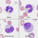
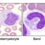
Immature neutrophils are classified based on their stage of maturation. The earliest identifiable neutrophil precursor is a myelocyte, which differentiates into a metamyelocyte, then a band neutrophil, and finally to a mature segmented neutrophil. Only the myelocyte is capable of division – all the more mature stages (metamyelocyte, band, segmented neutrophil) are incapable of division (post-mitotic). The primary criterion for differentiating immature neutrophils from each other is the shape of their nucleus, which starts to indent or constrict as the cell matures. A myelocyte has a round nucleus, a metamyelocyte has an indented or kidney-bean shaped nucleus and a band has a horse-shoe or parallel-sided shaped nucleus.
Immature neutrophils must be distinguished from monocytes, particularly when there is evidence of toxic change in the neutrophils. This can be difficult to do, but is usually accomplished by evaluating the entire cell (nuclear shape, nuclear location within cell, nuclear chromatin and cytoplasmic features). Immature neutrophils tend to show features of toxicity (cytoplasmic basophilia, Dohle bodies, cytoplasmic vacuolation), have more clumped chromatin than monocytes and when they are metamyelocyte or myelocyte forms, the nucleus is eccentric. Cytoplasmic borders, when they abut adjacent red blood cells tend to have a light rim. In contrast, monocytes have fatter nuclei which are more pleomorphic and tend to be more centrally located, they are somewhat larger than immature neutrophils and have a more uniformly colored blue-gray cytoplasm, which may contain small discrete-margined vacuoles. Streaky irregularity to the cytoplasm, which signifies toxic change in neutrophils, is generally not seen in monocytes. In addition, when monocytes (or lymphocytes) abut adjacent red blood cells, the rim of cytoplasm is dark. Refer to the hematology atlas for images showing the difference between monocytes and immature neutrophils.
Related links
- Interpretation of leukogram patterns
- Toxic change in neutrophils: Morphologic features
- Normal leukocytes: Morphologic features, including distinguishing bands from monocytes
- Hematology atlas: Atlas of images from different species, reflecting normal and abnormal findings.
- Historical origin of the term left shift