
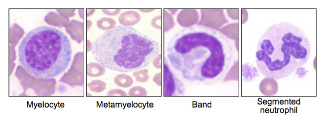
This image shows different stages of neutrophil maturation. They are taken from different dogs. Maturation proceeds from left to right, with the most immature stage in this image being the myelocyte with a round eccentric nucleus, followed by the metamyelocyte with a reniform (kidney bean-shaped) eccentric nucleus and a band neutrophil, with a horse-shoe shaped nucleus. The final image is the mature segmented neutrophil. All of the immature stages are toxic as seen by streaky blue cytoplasm (cytoplasmic basophilia), cytoplasmic rarefaction or indistinct vacuolation (most apparent in the metamyelocyte and band neutrophil) and lightening of the chromatin (less clumped than the segmented cell). The segmented neutrophil is not toxic. The myelocyte resembles a reactive lymphocyte, but the cytoplasm is too irregular and distinct aggregates of blue are seen at the edges of the cytoplasm, which are Dohle bodies and the chromatin is still coarsely clumped. The immature neutrophils are deliberately depicted to the left of the mature neutrophils (in sequential order of decreasing maturity) to illustrate the use of the term “left shift”.