
Peritoneal fluid from a dog
Case information
An 8 year old male castrated Chihuahua was presented to the Cornell University Hospital for Animals (CUHA) Emergency Service for evaluation of increased respiratory rate and effort. The owners reported that the day prior to presentation, the dog was lethargic and had vomited. The owners brought the dog to their primary veterinarian who noted a two pound weight loss since the last wellness exam (7 months prior), and performed routine bloodwork and two-view abdominal radiographs. The liver enzymes were reportedly markedly increased and the abdominal radiographs were unremarkable. The dog was discharged with metronidazole, Denamarin, and Fortiflora. The lethargy worsened and the dog developed hematochezia and respiratory difficulties, which prompted the owners to bring the animal to CUHA for further evaluation.
On presentation, the dog was bright, alert, and responsive, but was mildly febrile (103.2 F), tachycardic (170 bpm), and tachypneic. Other physical examination abnormalities included cranial organomegaly, a tense abdomen on palpation, and mucoid feces. A targeted ultrasound detected a small amount of free fluid in the peritoneal cavity. An abdominocentesis was performed and a sample was submitted for cytologic evaluation the next morning, along with a CBC, chemistry panel, and urinalysis. The CBC revealed a moderate leukocytosis (29,500/μL, reference interval [RI]: 5,700-14,200/μL) characterized by a mature neutrophilia (26,300/μL, RI: 2,700-9,400/μL) and monocytosis (2,400/μL, RI: 100-1,300/μL) with a mild lymphopenia (600/μL, RI: 900-4,700/μL). Abnormal chemistry results were as follows:
| Analyte | Result | RI | Units |
| Potassium | 3.8 | 4.1-5.6 | mEq/L |
| Creatinine | 0.4 | 0.6-1.4 | mg/dL |
| ALT | 377 | 27-98 | U/L |
| AST | 108 | 14-51 | U/L |
| ALP | 1960 | 17-111 | U/L |
| GGT | 21 | 0-6 | U/L |
| Total bilirubin | 0.4 | 0-0.2 | mg/dL |
| Direct bilirubin | 0.2 | 0-0.1 | mg/dL |
| Creatine kinase | 459 | 48-261 | U/L |
| Iron | 56 | 78-214 | μg/dL |
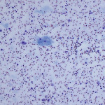
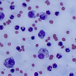
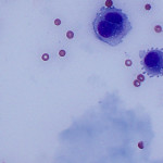
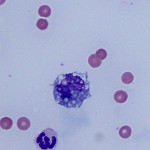
The submitted peritoneal fluid was dark red and opaque with a total protein by refractometer of 6.2 g/dL, nucleated cell count of 89.0 thou/μL, and RBC count of 651.0 thou/μL. Direct smears were prepared from the submitted fluid. Evaluate the chemistry results and representative images of the peritoneal fluid below, and consider the following questions:
- Based on the CBC and chemistry panel, what pathologic processes are present?
- How would you classify the effusion?
- What are your differential diagnoses for the underlying cause of the effusion?
- What additional diagnostic tests are indicated in this case?
|
|
|
|
|
Answer on next page