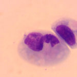
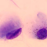
Conjunctival swab from a dog
Case information
A 3-month-old neutered male Siberian Husky puppy presented to the referring veterinarian with a 5 week history of respiratory symptoms and rhythmic jaw movements. An in-clinic hemogram revealed the following:
| Table 1: Abbreviated hemogram results | |||
| Test | Results | Units | Reference interval |
| HCT | 26 | % | 37-62 |
| MCV | 55 | fL | 62-74 |
| MCHC | 38 | g/dL | 32-38 |
| Retic | 0.5 | % | Not provided |
| Abs Retic | 24 | thou/μL | 10-110 |
| WBC | 2.4 | thou/μL | 5.1-16.8 |
| Seg Neut | 0.8 | thou/μL | 3.0-11.6 |
| Lymph | 1.1 | thou/μL | 1.1-5.4 |
| Platelets | 164 | thou/μL | 148-484 |
Results for an in-house biochemistry panel were within provided reference intervals.
The veterinarian treated the puppy for a presumptive pneumonia with antibiotics, but there was little improvement. The dog was subsequently admitted to an emergency clinic for anorexia and dyspnea. When examined at the emergency clinic, the puppy was dyspneic (this was oxygen dependent), with a marginal increase in temperature (102.6ºF), and had a bilateral serous nasal discharge and chemosis. A swab of the dog’s conjunctival mucosa was taken and rolled onto a microscope slide, followed by rapid staining (Diff-quik®).
Evaluate the representative photomicrograph of the conjunctival swab and answer the questions posed below:
- Are any abnormalities evident in the conjunctival swab?
- Do the observed findings explain the hemogram results?
- What are potential explanations for the dog’s microcytosis?
 |
 |
Answer on next page
