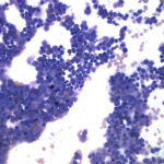
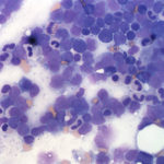
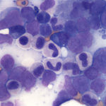
Bone marrow aspirate from a dog
Case information
A 5 year old, male castrated, Australian shepherd dog was referred for thrombocytopenia, hypercalcemia and peripheral lymphadenopathy. The dog also demonstrated a decreased appetite and energy level. Mild, intermittent diarrhea, as well as polyuria and polydipsia, were noted. Prior to the onset of these symptoms, the dog was considered healthy and active.
Upon presentation, the dog was bright, alert and responsive and vital parameters were within normal limits. The left superficial, cervical and submandibular lymph nodes were notably enlarged, with other peripheral lymph nodes being mildly increased in size. The remainder of the physical examination was within normal limits. Significant laboratory abnormalities included a moderate increase in total calcium [13.5 mg/dL, reference interval (RI) 9.3-11.4 mg/dL], with a concurrent marked increase in ionized calcium (1.65 mmol/L, RI: 1.18-1.37 mmol/L) and a marked thrombocytopenia (25,000/uL, RI: 186,000-545,000/uL) with no platelet clumping noted on review of the blood film. Low numbers of suspected circulating neoplastic cells, with morphologic features most suggestive of lymphoid origin, were also seen on blood film review. In addition, some atypical morphological features of neutrophils were noted. Thoracic radiographs revealed a possible sternal and tracheobronchial lymphadenopathy. Fine needle aspiration of peripheral lymph nodes was performed (the cytologic results will be discussed in the Explanation section). Bone marrow aspiration was also performed.
Evaluate the representative images of the bone marrow below and answer the following questions:
- What is the major process occurring in the bone marrow?
- Why is the dog thrombocytopenic?
- What additional diagnostic test could you perform to refine your diagnosis of the underlying process in this case?
- What morphological abnormalities do you detect in the neutrophils within the marrow sample? What is the significance of this finding?
|
|
|
|