Joint aspirate from a tortoise
Case information:
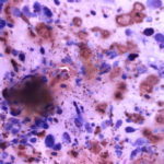
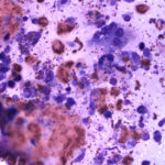
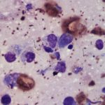
An approximately 18 month old, male Hermann’s tortoise presented for raspy breathing, exuberant, pink tissue over the eyes and being less active than normal. On physical examination the tortoise was small for his age, had increased respiratory rate and effort, was 5% dehydrated and would not open his eyes. While the tortoise was fully ambulatory, his left rear leg was swollen in the tarsal region. Whole body radiographs were taken to better assess the animal’s overall health. No abnormalities were seen in the lungs, but extensive bone loss and soft tissue swelling was noted in the region of the left tarsus. The left tarsal joint was aspirated and thick, gritty material was obtained. A portion of this sample was submitted for culture, while the remainder of the sample was assessed cytologically. Evaluate the photomicrographs of the submitted joint material and consider the following questions:
Questions:
- What types of inflammatory cells are present?
- What are the structures indicated by the arrows in Figure 3 (also pictured in Figure 1A)?
- What is the final diagnosis based on the cytological findings?


