
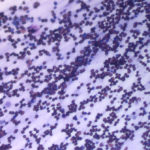
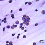
Tracheal wash from a horse
Case information
An 8 year old Morgan gelding presented to the Cornell University Hospital for Animals with a 3 week history of serohemorrhagic skin lesions and a 1 week history of a progressively worsening cough. The horse had also recently lost 100 lb. On physical examination, multifocal oozing lesions were present on the left hip, lower aspects of three limbs and neck. No abnormal lung sounds were detected on auscultation with or without a rebreathing bag. Thoracic ultrasonographic and radiographic examination revealed diffuse lung consolidation and pleuritis, with a mixed pulmonary pattern and coalescing nodules, respectively. Blood samples were taken for a hemogram plus fibrinogen by heat precipitation and chemistry profile.
Hemogram abnormalities included a mild neutrophilia of 7.3 thou/uL (reference interval: 2.7–6.6 thou/uL) and a mild hyperfibrinogenemia of 400 mg/dL (reference interval, 0-200 mg/dL). There was a mild hypoalbuminemia (2.5 g/dL, reference interval: 3.0–3.7 g/dL) and hyperglycemia (141 mg/dL, 71-113 mg/dL) on the biochemical panel.
An endoscopic tracheal wash and blind bronchoalveolar lavage were performed and submitted for cytologic evaluation.
Evaluate the representative photomicrographs of the direct smear of the tracheal wash and answer the questions posed below:
- What cell types are present in the smears (how would you characterize the results)?
- What are your differential diagnoses for the findings?
|
|
|
Answer on next page