Peritoneal fluid from an alpaca
Case information
A 6 year old female alpaca presented to the Cornell University Large Animal Hospital with a history of recumbency, anorexia, and dull mentation. Initial physical examination and diagnostic testing showed the animal was dehydrated and had clinical signs consistent with a proximal intestinal obstruction (decreased fecal output, significantly distended third gastric compartment, empty small intestine, and a shadowing object in the caudal aspect of the duodenal ampulla on abdominal ultrasonography). The hemogram showed a mild lymphopenia of 0.2 thousand/µL (reference interval, 1.1-5.5 thousand/µL), likely due to stress. Pertinent biochemical results are shown on the table below:
| Analyte | Result | RI | Units |
| Sodium | 160 | 149-157 | mEq/L |
| Chloride | 106 | 106-116 | mEq/L |
| Bicarbonate | 41 | 22-34 | mEq/L |
| AST | 1062 | 119-286 | U/L |
| SDH | 117 | 0-7 | U/L |
| GLDH | 540 | 3-19 | U/L |
| GGT | 155 | 8-35 | U/L |
| Bilirubin | 0.1 | 0-0.1 | mg/dL |
The patient was taken to surgery for duodenal enterotomy and third gastric compartment gastrotomy. Surgery revealed two bezoars, one partially obstructing the distal third gastric compartment and another obstructing the duodenal ampulla. An increased amount of peritoneal fluid was found adjacent to the obstructed duodenum. The bezoars were surgically removed and a sample of peritoneal fluid was collected and submitted for cytologic evaluation.
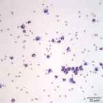
The submitted peritoneal fluid was yellow to light-red in color and opaque, with total protein by refractometry of 2.7 g/dL. The total nucleated cell count was 6.5 thousand/µL and a red blood cell count was 65.1 thousand/µL. Direct and sediment smears from the submitted fluid were prepared.
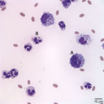
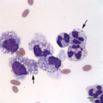
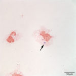
Evaluate the representative photomicrographs of the peritoneal fluid shown below, consider the biochemistry results, and answer the following questions:
- What type of metabolic derangement is present?
- Identify and evaluate the cells labeled as “A” and “B” in Figure 2.
- What additional diagnostic tests would you do in this case?
|
|
|
|
|