
Fever of unknown origin in a 7-month-old cat
Case Information
A 6 lb intact male Domestic Medium hair cat estimated to be 7 months old was presented to Cornell’s Emergency Service with an acute onset of lethargy. The cat was one of the kittens born in a window well at the owner’s house and had been living outdoors in a wooded area. The cat was found lying still, lifting its head occasionally and walking a few steps. The cat had not been vaccinated and the owner reported observing many ticks in the past. A topical preventative containing fipronil and (S)-methoprene was administered the day before presentation.
On physical examination, the cat was bright, alert, and responsive. Severe pyrexia (107.1 F), tachycardia (240 beats per minute), tachypnea (66 breaths per minute), and an estimated 5% dehydration were noted. Initial point-of-care bloodwork revealed a packed cell volume of 34% (reference interval [RI]: 31-48%), increased total solids of 8 g/dL (RI: 5.9-7.5 g/dL) and a mild hyperglycemia of 146 mg/dL (RI: 63-140 mg/dL). An azostix was normal at 5-15 mg/dL. Blood gas analysis showed a low normal pH (7.33 units, RI: 7.32-7.3.8 units) with a metabolic acidosis (bicarbonate, 14 mmol/L, RI: 20-25 mmol/L; base excess, -11 mmol/L, RI: 0 to -4 mmol/L) and a normal anion gap (19 mmol/L, RI: 14-29 mmol/L) with a concurrent respiratory alkalosis (pCO2, 29 mmHg, RI: 38-46 mmHg). Electrolyte testing showed a hyponatremia (143 mEq/L, 149-157 mEq/L) and hypochloremia (114 mEq/L, corrected chloride, 122 mEq/L, RI: 117-127 mEq/L). A rapid test for Dirofilaria immitis and feline leukemia virus antigens and feline immunodeficiency virus antibodies was negative. A fast assessment abdominal ultrasonographic examination revealed no significant abnormalities. Thoracic radiographs revealed a mild, diffuse bronchial pattern. The cat was hospitalized and given intravenous fluids (Plasmalyte) and anti-emetics. The next morning, blood and a urine sample collected by cystocentesis were submitted to the Clinical Pathology laboratory for a hemogram, biochemical profile and urinalysis, respectively. By that time, the cat’s fever had decreased to 105 F and an ocular discharge was noted.
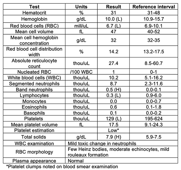
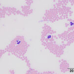
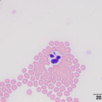
Biochemical panel results revealed an ongoing mild hyponatremia (144 mEq/L; RI:149-158 mEq/L) and hypochloremia (110 mEq/L, corrected chloride of 117 mEq/L, RI: 114-124 mEq/L). Urea nitrogen (10 mg/dL; RI: 17-35 mg/dL) and creatinine (0.7 mg/dL; RI: 0.8-2.1 mg/dL) concentrations were decreased and there was a mild hyperphosphatemia (6.0 mg/dL; RI: 2.6-5.5 mg/dL). A mild hypoferremia (16 ug/dL; RI: 20-56 ug/dL) with low transferrin saturation (6%; RI: 20-46%) was present. Urinalysis results showed a trace protein on a dipstick, with a urine specific gravity of 1.025, but were otherwise unremarkable. The hemogram results and representative images of the blood smear are shown below:
|
|
|
- Do you see a cause for the cat’s clinical signs in the blood smear?
- What additional tests would you perform?
Answers on next page