Blood smear and peritoneal fluid from a cat
Case information:
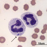
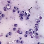
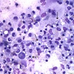
A 4 year old male castrated domestic shorthair cat presented with a 4 day history of anorexia, lethargy and vomiting. The referring veterinarian had administered Convenia and Metacam, with no improvement noted by the owner. Upon presentation to the Cornell University Hospital for Animals, the cat was laterally recumbent, depressed, and dehydrated (estimated at 7-10%). The cat had a body condition score of 8/9 (obese). Blood was drawn for a CBC and full chemistry panel. The CBC revealed a moderate anemia with minimal regeneration and degenerative left shift with a normal neutrophil count and marked toxic change (Figure 1A). Abdominal tension and mild pain on palpation was noted. Abdominal ultrasound revealed effusion and raised concern for foreign bodies in the stomach and a suspect linear foreign body in the intestine. Peritoneal fluid was collected and submitted for cytological examination and bacterial culture. The nucleated cell count of the fluid was 2,600 cells/ul and the protein concentration was 3.5 g/dL. Lactate and glucose were also measured on the peritoneal fluid sample, and were 2.6 mmol/L and 302 mg/dL, respectively. Abbreviated chemistry results are listed below, as well as photomicrographs of the blood smear and peritoneal fluid (Figures 1 B and 1C). The urinalysis was unremarkable.
Abbreviated biochemistry results:
| Analyte: | Value: | Reference Range: |
| Sodium | 131 mEq/L | 151-158 mEq/L |
| Chloride | 91 mEq/L | 113-123 mEq/L |
| Calcium | 7.1 g/dL | 9.1-10.9 g/dL |
| Glucose | 315 mg/dL | 64-144 g/dL |
| Alk Phos | 5 U/L | 13-83 U/L |
 |
 |
 |
Questions:
- Is there biochemical evidence that supports on-going vomiting?
- How would you classify the cat’s peritoneal fluid?
- Are the cytological findings consistent with a perforated GI tract and septic peritonitis?
- What material do you suspect is in the macrophages (Figure 1B, long arrow) and neutrophils (arrows in Figure 1C)?
Answer on next page
