
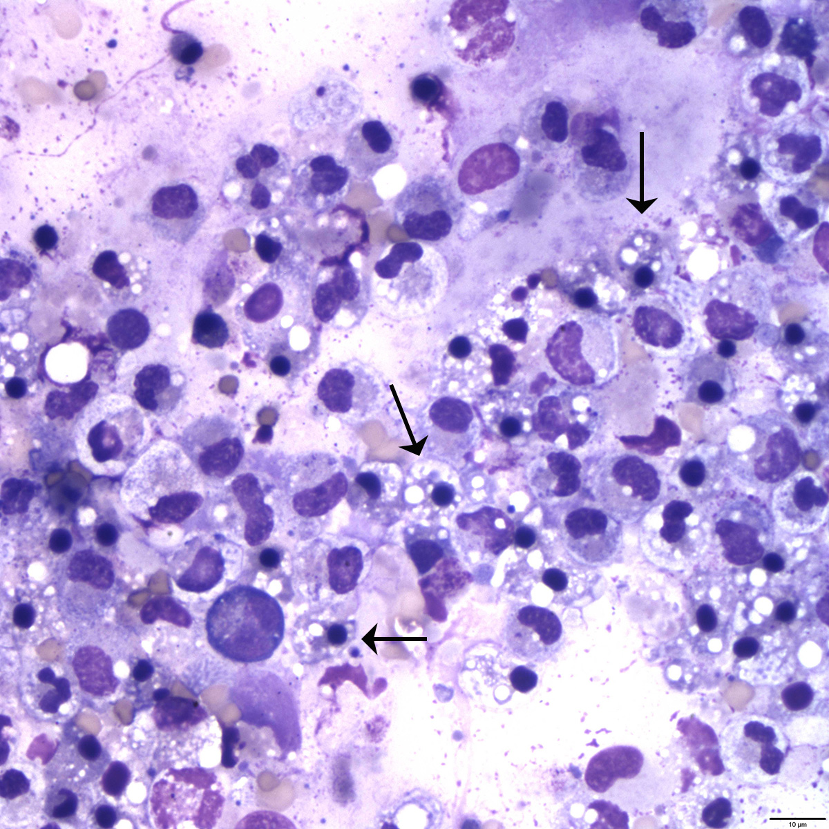
Photomicrograph of peritoneal fluid. Note the abundant numbers of pyknotic cells indicated by the arrows. Cytocentrifuged sample, Wright’s stain, 50X.
Photomicrograph of peritoneal fluid. Note the abundant numbers of pyknotic cells indicated by the arrows. Cytocentrifuged sample, Wright’s stain, 50X.
eClinpath helped 1.2 million visitors last year from 220 countries find important information on animal health. If you enjoy the site, please support our mission and consider a small gift to help us keep pace with its rapid growth. You can donate securely via PayPal or credit card. Thank you!
![]()