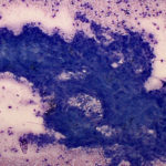
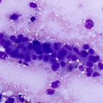
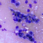
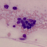
Liver imprint from a horse
Case information
An 11-month-old Thoroughbred filly presented to the Cornell University Hospital for Animals with a 10 day history of persistent lethargy, intermittent fever, diarrhea and hypodipsia. There was no response to antibiotics and fluids administered by the referring veterinarian.
On physical examination, the filly was quiet, alert and responsive, but in poor condition with a distended “pot-bellied” abdomen and dehydration. Mucous membranes of the oral and vaginal mucosa were hyperemic and the capillary refill time was 3 seconds. There were decreased borborygmi on abdominal auscultation. The filly had high heart and respiratory rates, with increased bronchovesicular sounds, and was afebrile.
Ultrasonographic examination of the thorax and abdomen revealed mild pleural roughening and a markedly enlarged liver that was comprised of several large or multiple coalescing masses, with intraluminal material in portal and splenic veins (suspected thrombosis). A 21 x 25 cm complex mass with a liquid center was noted in the cranial part of the abdomen. Thoracic radiographs did not reveal any abnormalities. Point-of-care testing revealed a packed cell volume of 64% and total protein by refractometer of 8.4 g/dL.
The filly was administered a bolus of 15 liters of intravenous fluids and then was treated with antibiotics and intravenous fluids overnight. The next day, blood was sampled for hematologic and biochemical testing (Tables 1 and 2) and a screening coagulation panel. Coagulation testing revealed a prolonged prothrombin time (27 seconds, reference interval 16-20 seconds) with a normal activated partial thromboplastin time (59 seconds, reference interval 45-66 seconds) and hyperfibrinogenemia (1131 mg/dL, reference interval 175-445 mg/dL). Bile acid testing revealed a high normal concentration (11 μmol/L, reference interval 0-11 μmol/L).
| Table 1: Pertinent hematologic results | |||
| Test | Result | Units | Reference interval |
| Hematocrit | 51 | % | 31-47 |
| MCV | 33 | fL | 38-50 |
| White blood cells | 7.6 | thou/μL | 5.5-11.4 |
| Neutrophils | 4.6 | thou/μL | 2.7-6.6 |
| Platelets | 437 | thou/μL | 98-246 |
| MPV | 7.6 | fL | 5.8-11.5 |
| Total protein by refractometer | 8.9 | g/dL | 5.2-7.8 |
| Table 2: Pertinent biochemical results | |||
| Test | Result | Units | Reference interval |
| Sodium | 129 | mEq/L | 134-141 |
| Chloride | 94 | mEq/L | 95-106 |
| Bicarbonate | 22 | mEq/L | 24-31 |
| Anion gap | 18 | mEq/L | 8-19 |
| Albumin | 2.1 | g/dL | 2.8-3.8 |
| Globulin | 5.7 | g/dL | 2.4-4.4 |
| A/G | 0.37 | 0.8-1.4 | |
| Glucose | 52 | mg/dL | 75-117 |
| AST | 485 | U/L | 212-426 |
| SDH | 6 | U/L | 2-11 |
| GGT | 128 | U/L | 8-33 |
| Total bilirubin | 4.0 | mg/dL | 0.8-2.2 |
| Direct bilirubin | 0.5 | mg/dL | 0.1-0.3 |
| Indirect bilirubin | 3.5 | mg/dL | 0.5-2.1 |
| CK | 112 | U/L | 93-348 |
| Iron | 67 | μg/dL | 105-277 |
| Transferrin saturation | 21 | % | 26-62 |
The hepatic mass was biopsied under ultrasonographic guidance and scrapings were made from the submitted tissue, smeared onto slides and stained with modified Wright’s stain. Examine the representative images of the smears, then answer the questions below:
- Do most of the cells in the aspirate have features typical of mature hepatocytes?
- What is your cytologic diagnosis, incorporating all observed cytologic findings?
- Does this diagnosis provide pathophysiologic mechanisms that would explain some of the hematologic and biochemical findings?
 |
 |
 |
 |
Answers on next page
