“Pulmonary mass” in a dog
Case Information
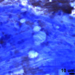
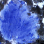
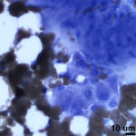
A 6 year old mixed breed dog presented for difficulty breathing. A single pulmonary mass was identified, and a fine-needle aspirate collected and submitted for cytological evaluation. CBC and biochemical panel results were unremarkable. Images of the aspirate are shown below:
Questions:
1) What is the major cell type seen in this aspirate?
2) Take a close look at image 4, what second population of cells is evident?
3) How do you reconcile these findings with a lesion in the thoracic cavity?


