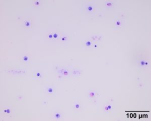
Examination of a direct smear of the fluid at low magnification revealed a predominance of macrophages/synoviocytes with lower numbers of eosinophils and small lymphocytes. The background contains a few erythrocytes and free eosinophil granules. The purple and stippled background is a common finding in joint fluid as the fluid contains proteins that yields the characteristic viscosity (e.g. hyaluronic acid) (Wright’s stain, 20x objective).

