
Synovial fluid from a horse
Case Information
A 13 year old Draft mix gelding presented with a 2 week history of left front limb lameness. On physical examination, there was moderate joint effusion of the fetlock joint.
Synovial fluid was aseptically collected into a plain red top tube (without anticoagulant) and a purple top tube (with EDTA anticoagulant) and submitted for fluid analysis and culture to the Animal Health Diagnostic Center (AHDC) at Cornell University. Prior to submission, direct smears from the freshly collected fluid were made and submitted along with the fluid. The results of the fluid analysis are provided below.
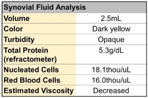
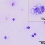
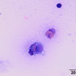
Provided below are representative images of the joint fluid.
|
|
|
|
")
Using the provided information, answer the following questions;
- What are the cell populations in the fluid and which population is dominating?
- How would you interpret the joint fluid based on the nucleated cell count, total protein concentration and the provided images?
- What would be your differential diagnoses?
Answers on the next page.