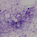
Photomicrographs of cytology of a dried fecal smear from a puppy
Case Information
A 3-month old, female, mixed breed puppy was presented to the Cornell University Hospital for Animals for evaluation of chronic diarrhea. She had recently been adopted from a local shelter. She was part of a litter of 3 puppies. Reportedly, one of the puppies died at 8 weeks of age from a presumed canine parvovirus infection. The other puppies had milder symptoms, and had responded to symptomatic treatment. On physical examination, the puppy had mild vaginitis, but appeared otherwise healthy. Cytology of a dried fecal smear was performed.
Evaluate the representative photomicrograph of the dried fecal smear cytology (Figure 1) and answer the following questions:
1. What is the nature of the organisms and structures present in the fecal smear?
2. What are your differential diagnoses for the larger structures based on the images?
3. What additional diagnostic tests would you recommend?
 |
Answer on next page
