
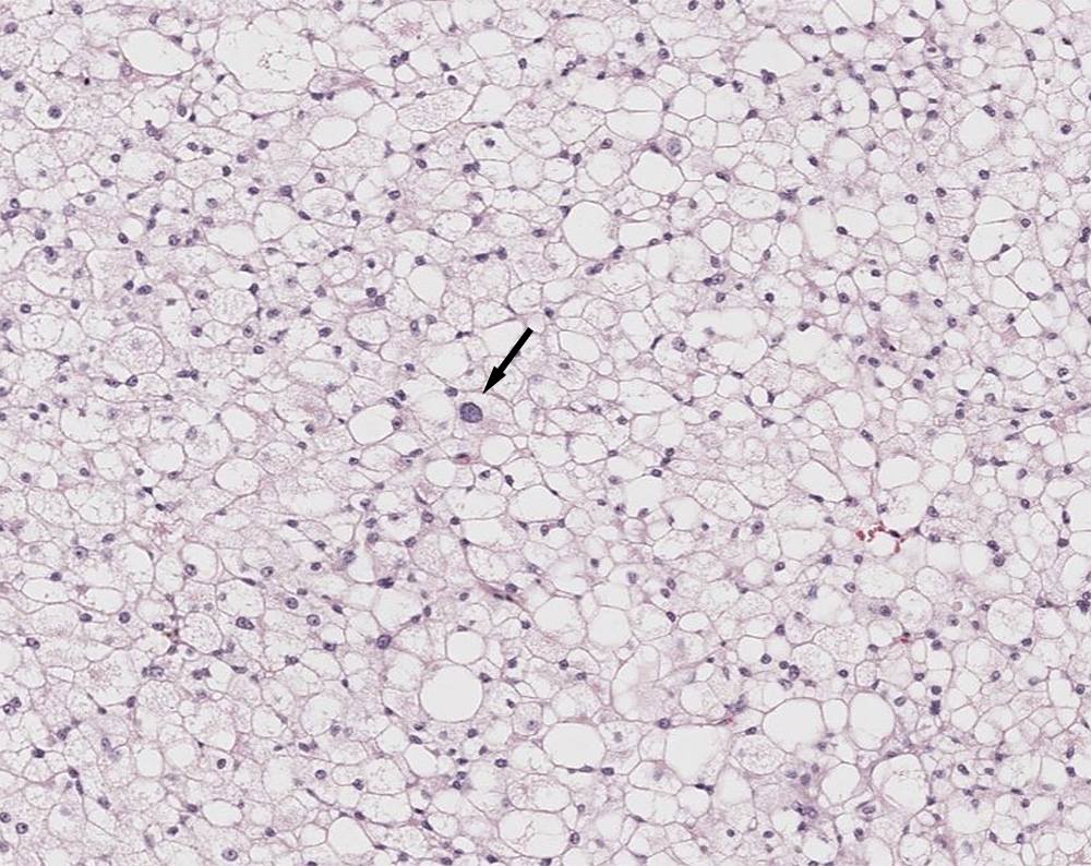
The biopsy consisted of sheets of round to polygonal cells with abundant vacuolated to slightly pink grainy cytoplasm, which are characteristic of brown fat (pale cells). Several cells have single large cytoplasmic vacuoles with eccentric nuclei, resembling adipocytes. Anisokaryosis and anisocytosis were mild overall, although individualized cells with larger nuclei were seen (arrow). The histologic diagnosis was a hibernoma, based on the mixture of mostly highly vacuolated cells and fewer cells with a single large lipid droplet (hematoxylin & eosin stain).