
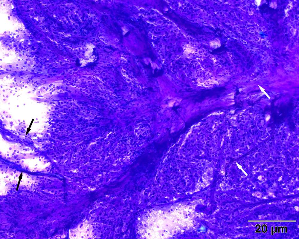
Numerous vascular profiles (arrows) are seen coursing through the mass with affiliated tumor cells. Diff-quik stain, 10x objective
Numerous vascular profiles (arrows) are seen coursing through the mass with affiliated tumor cells. Diff-quik stain, 10x objective
eClinpath helped 1.2 million visitors last year from 220 countries find important information on animal health. If you enjoy the site, please support our mission and consider a small gift to help us keep pace with its rapid growth. You can donate securely via PayPal or credit card. Thank you!
![]()