
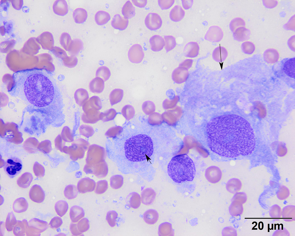
Higher power view of the tumor cells, showing their spindled outlines (long arrow) and indistinct nucleoli (short arrow). These cells display moderate anisokaryosis and anisocytosis (modified Wright’s stain)
Higher power view of the tumor cells, showing their spindled outlines (long arrow) and indistinct nucleoli (short arrow). These cells display moderate anisokaryosis and anisocytosis (modified Wright’s stain)
eClinpath helped 1.2 million visitors last year from 220 countries find important information on animal health. If you enjoy the site, please support our mission and consider a small gift to help us keep pace with its rapid growth. You can donate securely via PayPal or credit card. Thank you!
![]()