
Aspirate of a retrobulbar mass in a dog
Case Information
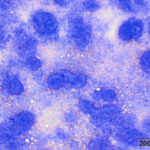
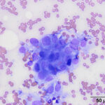
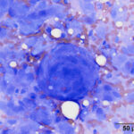
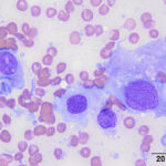
A 6-year-old male neutered mixed breed dog was presented to the Ophthalmology Service at Cornell University for evaluation of an optic nerve tumor in the right eye. Approximately one year prior, the patient had developed a raised third eyelid and exophthalmos in the right eye, which ultimately progressed to vision loss. On physical examination, the patient’s right eye was exophthalmic with scleral injection. The pupillary light reflex and menace response were absent. Upon fundic examination, retinal detachment and increased size of the optic nerve were noted. A computerized tomography (CT) scan of the skull revealed a large retrobulbar mass with intraocular and intracranial extension and a separate discrete intracranial nodule. Fine needle aspirates were taken from the retrobulbar mass for cytologic examination.
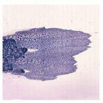
Examine the representative images of the smears or view the digital slide by clicking on the image to the right and answer the questions below:
- Based on the location, pattern of cell arrangement and shape of the cells, what is your primary differential diagnosis?
- What additional tests can be performed to confirm your diagnosis?
|
|
|
|
|
Answers on next page