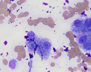
Dark blue granules of presumptive copper are seen in these well-spread hepatocytes (the long arrow indicates the pigment in one of the cells). Note the abundant bright magenta ultrasound gel in the background and between hepatocytes (short arrow). This gel obscured many of the cells (modified Wright’s stain, 100x objective).

