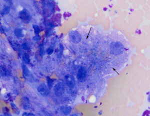
The hepatocytes on the edge of this dense cluster are slightly distended and contain moderate amounts of blue-green granules in the cytoplasm (arrows). Rare granules were more turquoise and slightly crystalline when the focus was moved up and down. This is hard to capture in a still image (modified Wright’s stain, 100x objective). The latter color and texture is typical of copper, however the other pigment can be (and was initially) misidentified as lipofuscin.

