Fat
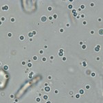
Variably-sized droplets of free lipid are fairly commonly observed upon microscopic examination of urine from dogs and cats.
They can most readily be differentiated from cells by their propensity to float up under the coverslip to a different plane of focus compared to cells which have settled to the surface of the slide. They are uniformly round, but more variable in size than cells and are smooth in appearance. By focusing up and down, one can also visualize their perfectly round (droplet) appearance, even when suspended in urine.
Urine lipid originates mainly from degeneration (physiologic or pathologic) of epithelial cells lining the urinary tract. As an isolated finding, it is often not of clinical significance. There is no clear association with hyperlipidemia.
Mucus
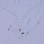
Mucus can be observed in some urine samples, particularly that collected from horses (hence it is a constituent and not a contaminant). Mucus is far more infrequent in urine samples from other species.
Strands of mucus can mimic hyaline casts, particularly when viewed under regular light microscopy.
Compared to casts: Mucus is more irregular in shape and has irregular borders with tapered ends.
Contaminants
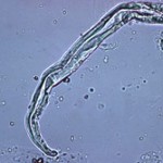
Extraneous contaminating materials of many types can make their way into urine specimens. Generally, such material is significant only as an indication of the conditions of collection and/or handling of the specimen prior to analysis. Debris is more frequently seen in voided urine samples or those collected off the floor, from litter trays and when in an environment such as a courtyard or barn. On occasion, confusion may arise if these are mistaken for findings of significance (casts, parasites, etc.).
Striving for optimal collection and transport of specimens will help maximize useful results and minimize confusing findings. Specimens mailed to laboratories without refrigeration or preservatives are subject to microbial overgrowth, whether contaminants or pathogens. Refrigeration is perhaps the best all-around method for preserving a specimen.
Fecal parasites
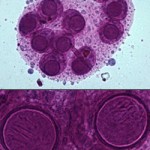
Fecal material, including eggs of intestinal parasites, can contaminate voided urine samples. For example, the images on the right show a urine sediment of a cat stained with Sedi-stain. In the upper panel is a low magnification view showing a packet of eggs. In the lower panel, higher magnification reveals the internal structure of the hexacanth embryos of Dipylidium caninum, a tapeworm.
Environmental fungi
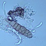
Mold spores come in a variety of shapes, sizes and colors.
Shown on the right is a brownish Alternaria spore surrounded by amorphous crystals and a few lipid droplets. Alternaria is a dematiaceous fungus that is ubiquitous in the environment.
Fibres
Cotton, plant, and paper fibers may mimic parasite larvae or urinary casts
Pollen
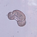
Pollen grains are generally round to oval and some have a yellow to brown tint. They are most likely to be confused for parasitic ova. A good knowledge of the actual appearance of the few true parasite eggs that can occur in urine is easier to achieve than specific recognition of all types of pollen and mold spores. Again, optimizing specimen collection and handling will reduce the chances of seeing potentially confusing structures.
Starch crystals
Starch crystals are typically from lubricant in surgical and examination gloves. They are common contaminants in urine sediments and cytology smears.
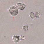
They are usually variable in size, round to polygonal in shape, colorless, refractile (due to their crystalline nature) and usually have a circular or Y-shaped “dot” in the center (arrow in image on right).
Microbial overgrowth
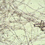
Shown here is a dense mat of fungal hyphae in canine urine which was several days in transit. Since fungal infection of the urinary tract in dogs is quite uncommon, the odds are that this represents overgrowth of contaminants.
Bacteria, whether pathogens or contaminants, can multiply when analysis is delayed. This often clouds interpretation of the sediment examination and culture results.
