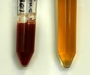
The urine on the left is dark red. On centrifugation, the pellet was red and the supernatant was light yellow and non-turbid (clear). Numerous RBC (>100/HPF) were seen in the wet preparation of the urine when examined microscopically. This indicates a gross hematuria (macroscopic). A dipstick showed a 4+ heme reaction due to lysis of the RBC as they contacted the pad. In contrast, the urine on the right is medium yellow, which is normal for concentrated urine. Although there is no hint of red, it is still possible that a mild microscopic hematuria is present. However, the heme reaction on the dipstick was negative and a sediment examination revealed no RBC/HPF, which is normal (i.e. no microscopic hematuria).

