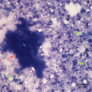
The aspirate revealed clusters of disorganized epithelial cells (orange arrow) immersed on a purple stippled background with many keratinized squamous epithelial cells (green arrows) individualized throughout the sample (Wright’s stain, 20x objective).

