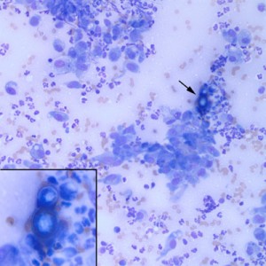
Figure 2: A single Blastomyces dermatitidis yeast is present in the upper right corner of the photomicrograph (arrow, Wright’s stain, x20 objective). Inset: Higher magnification of the same yeast organism. Note the thick double-contoured wall and broad-based budding (x50 objective).

