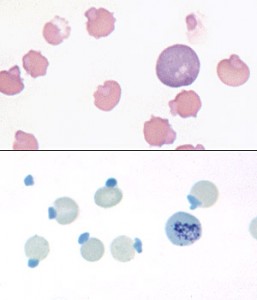
Blood from a cat with Heinz body hemolytic anemia associated with acetaminophen toxicosis.
Upper panel:the large Heinz bodies are causing severe distortion of the cell outline. Note the eccentrocyte in the upper left corner, and the ghost red cell with attached Heinz body in the upper right corner. the anemia is regenerative as there is a polychromatophil present (which corresponds to an aggregate reticulocyte).
Lower panel: Heinz bodies are readily visualized if the blood is incubated for several minutes in equal volume with New Methylene Blue. Heinz bodies take up the stain and are seen as blue inclusions on the edge of the cell. An aggregate reticulocyte is also present in this image. Note, that Howell-Jolly bodies will also stain blue with this stain and are difficult to differentiate from Heinz bodies (Howell-Jolly bodies should stain darker, more regularly round, and will not be protruding from the surface of the cell, as seen with these Heinz bodies).

