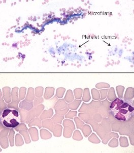
These pictures show the low and high power appearance of the feather edge, which is too thin to use for identification of leukocytes and assessment of morphologic too thin abnormalities. This part of the smear should be scanned at low power to detect platelet clumps and microfilaria, as shown in the top panel, but should be avoided when using the oil immersion objective. Abnormally large cells of potential diagnostic importance also tend to be drawn to the end of the smear

