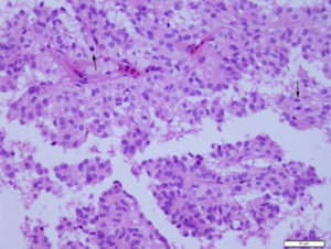
In some areas of the mass, the cells formed indistinct islands or lobules comprised of multiple disorganized layers of cells. The cells were polygonal to columnar and had pink lightly grainy cytoplasm with central to eccentric nuclei containing coarsely stippled chromatin with a single nucleolus. There are several mitotic figures (arrows) in this field. In this image, they show mild to moderate anisocytosis and anisokaryosis (mostly mild) (H&E, 40x objective). A Fite Faraco stain did not reveal any bacteria.

