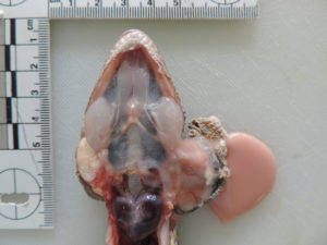
When sectioned, about 3 mls of pink white opaque fluid spilled from the sac. The larger mass was pedunculated and arising from the left lateral wall – it is visible in the upper part of the sac. A smaller mass was seen rostrally and on the medial wall of the sac. Histologic features of both masses were similar.

