Aspirate from a cervical swelling in a Tokay gecko (Gecko gecko)
Case Information
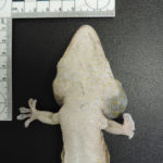
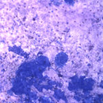
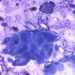
A captive 11 year old female Tokay gecko presented with a chronic (1 year) slowly progressive bilateral swelling of the upper cervical region (Figure 1). The swelling was more pronounced on the left side and had expanded more rapidly in the preceding 4 weeks. Other than size discrepancies, there were also differences noted between the two sides on palpation; the swelling was soft on the right side versus firm on the left side. Main findings on blood work were anemia (17% packed cell volume, average ÷ standard deviation reference value 30 ± 2% [n=6]1), hypercalcemia (48.4 mg/dL, average ÷ standard deviation reference value, 17.6 ± 0.4 mg/dL [n=2]1) and hypoalbuminemia (0.8 g/dL, reference animal [n=1], 2.7 g/dL1). On radiographs, both sides of the cervical region contained irregular mineralized opacities, however the left region also contained an enlarged diffuse soft tissue opacity with more irregular and lighter mineralized regions.2 The location of the mineralized areas on both sides was compatible with the endolymphatic sac. The swelling on the left cervical region was aspirated and yielded an opaque, white pink-tinged fluid. Removal of the fluid revealed a firm mass, which was also aspirated. Direct smears of the aspirated fluid and smears of the mass were examined and yielded similar cytologic findings (Figures 2-3).
Evaluate the provided cytologic images from the aspirated fluid, then answer the following questions:
- What cells and structures can be identified in the direct smears of the aspirates?
- What is your cytologic diagnosis?
- Do the cytologic results explain the abnormalities in the hematologic and biochemical results?
 |
 |
 |
Answers on the next page
