Ferritin is a large protein shell (MW 450,000, apoferritin) comprised of 24 subunits, covering an iron core containing up to 4000 atoms of iron. Apoferritin with bound iron (=ferritin) is the storage form of iron in cells and most readily available for use when needed. Ferritin is considered a better indicator of total body iron stores (non-heme iron) in animals than iron (which is a poor marker of total body iron stores). Serum ferritin concentrations are quite stable from day-to-day, in contrast to iron. Thus, measurement of ferritin can be used to confirm cases of iron deficiency or iron overload. However, the testing is not frequently performed because ferritin is influenced by factors other than body iron stores (see below) and the test is not offered by most veterinary diagnostic laboratories.
Physiology
Ferritin acts as the soluble storage form of iron in tissue (hemosiderin is relatively insoluble). It may serve other functions as well although these are controversial. It is found in most cells of the body, especially macrophages, hepatocytes and erythrocytes. Synthesis occurs in the liver and the rate of synthesis correlates directly with the cellular iron content. Control of ferritin synthesis occurs post-transcriptionally (at the mRNA level) and there are iron- and cytokine-responsive elements in ferritin mRNA. Thus ferritin levels change in response to iron stores and cytokine levels.
Methodology
Sensitive methods are needed, since serum levels are very low (in nanogram quantities). Immunologic assays requiring species-specific reagents, such as radioimmunoassay and a sandwich ELISA, have been employed. Canine and feline-specific ferritin assays are available through Kansas State University (see below).
Results are reported as ng/ml (conventional units) or pmol/L (SI units). The conversion factor is given below.
ng/mL x 2.247 = pmol/L
Test interpretation
Decreased concentrations (hypoferritinemia)
- Iron deficiency: A decrease in the amount of stored iron is the only known cause for a low serum ferritin result. This is the strong point of this test. However, in dogs or cats, serum ferritin values do not appear to correlate very well with non-heme iron stores in the liver. Indeed, the correlation coefficients between non-heme iron and ferritin are <0.4 in both species (Weeks et al., 1989, Andrews et al., 1994), indicating that other factors account for more than 60% or more of the variation in ferritin results. We have seen normal ferritin values in dogs with documented iron deficiency (absent bone marrow iron stores with accompanying microcytic hypochromic anemia). There is no apparent explanation for this, although these dogs could have concurrent inflammation (which does appear to be the case in some of these affected dogs). In cats, ferritin is anecdotally a better indicator of iron stores than in dogs.

Increased concentrations (hyperferritinemia)
In human patients, very high serum ferritin (>10,000 ng/mL) was associated with chronic transfusions, liver disease, cancer, macrophage disorders, and hereditary hemachromatosis (Sackett et al 2016).
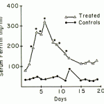
- Inflammation: Ferritin is an acute phase protein and values increase in response to inflammation. This is one of the causes of iron “sequestration” that occurs in animals with chronic or inflammatory disease and will reduce serum iron values. This limits the diagnostic use of this test for confirmation of iron deficiency anemia in dogs in particular.
- Macrophage disorders: Reactive conditions involving macrophages, e.g. hemophagocytic lymphohistiocytosis, can be associated with hyperferritinemia in humans. This has been attributed to high concentrations of inflammatory cytokines (Morimoto et al 2016).
- Neoplasia: Certain neoplasms, e.g. histiocytic sarcoma in dogs, are associated with high ferritin concentrations (Friedrichs et al 2010).
- Iron overload: Massive blood transfusions, hemochromatosis (rare).
- Liver disease: Liver disease, including acute hepatitis, non-alcoholic steatosis, viral hepatitis and cancer (e.g. hepatocellular carcinoma), in humans can be associated with high ferritin (Nakano et al 1984, Fargion et al 2001, Omagari et al 2003, Yenson et al 2008, Koto et al 2009). The high ferritin in hepatitis has been attributed to macrophage activation (Koto et al 2009) but it is possible that direct release of intracellular ferritin stores in necrotic hepatocytes is responsible. In one study, liver disease was the third most common cause of extremely high ferritin (following viral infections and hematopoietic neoplasia) (Sackett et al 2016).
Related links
- Kansas State University: Offers measurement of serum ferritin in various species, such as dogs, cats, rhinoceros, horse, dolphin.
