
Aspirate of a subconjunctival mass in a dog
Case Information
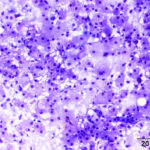
A 10-year-old female spayed mixed breed dog was presented to the Cornell University Hospital for Animals for a medial subconjunctival extraocular mass of the left eye. The mass was first noted approximately 3 months prior to presentation, when the owners observed hyperemia and serous discharge from the eye. The animal was treated by the primary veterinarian with topical antibiotics, non-steroid anti-inflammatory drugs, and a cefovezin (Convenia) injection. The dog was also reported to have ataxia and had a previous history of ear infections.
A complete blood count and biochemistry panel were performed, which were unremarkable. A computerized tomography scan of the head and neck showed a 1.6 cm in diameter, ovoid, well-demarcated, encapsulated mass medial to the left eye, between the globe and the orbital wall. A thin stalk at the caudal aspect of the mass extended into the rostral aspect of the medial rectus muscle. The right tympanic cavity was noted to be filled with fluid or soft tissue-attenuating material, which was interpreted as right otitis media, in line with the previous infections.
An abdominal ultrasound showed a few small hepatic and splenic nodules, which were interpreted as benign aging-related changes. A fine-needle aspirate of the subconjunctival mass was performed and submitted for cytologic examination.
View the provided images then answer the questions below.
 |
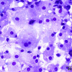 |
- What are your differential diagnoses based on the cytologic findings?
- Which additional test(s) would you run to further determine the origin of this mass?
Answers on next page