
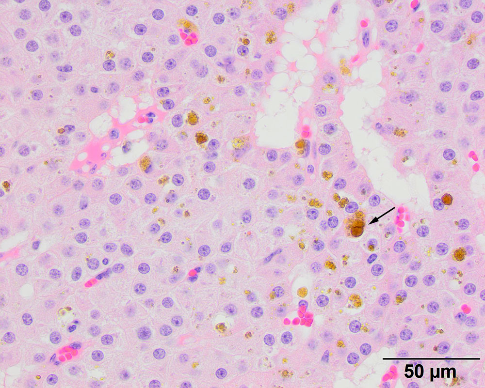
The neoplastic cells are polygonal to columnar (see cells on middle right) and display mild anisokaryosis and anisocytosis. They have pink grainy cytoplasm. Hemosiderophages are interspersed between the cells, indicating concurrent hemorrhage (hematoxylin-&-eosin, 20x objective). The histologic diagnosis was a parathyroid adenoma.