
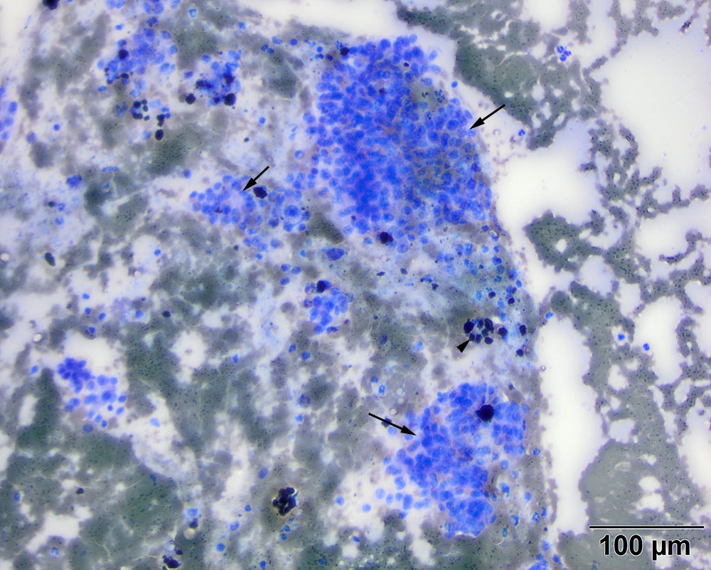
The aspirate consisted of numerous dense clusters (packets) of medium uniform epithelial cells (long arrows), with acinar-like arrangements (likely indicating follicular structures; short arrow). There were pigment-laden cells, compatible with hemosiderophages, indicating concurrent hemorrhage (arrowhead) (modified Wright’s stain, 20x objective)