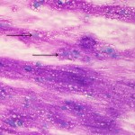
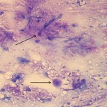
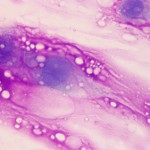
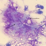
Photomicrographs of a fine needle aspirate from an epiglottic mass in a dog
Case information
An 11 year old male neutered Cavalier King Charles Spaniel presented for evaluation and treatment of periodontal disease. Upon intubation, a mass was observed arising from the dorsal surface of his epiglottis, extending laterally towards the right lateral commissure. The mass was described as firm. A fine needle aspirate was taken and the sample was submitted for cytologic evaluation. Photomicrographs of the aspirate are shown below. While the majority of the sample appeared similar to the area depicted in Figure 1, there were areas that had a more varied appearance (Figure 2).
- What are your differential diagnoses for the magenta and basophilic material dispersed throughout the background of the specimen?
- What are your differential diagnoses for the types of tumors that can arise in laryngeal tissue?
 |
 |
 |
 |
Answer on next page
