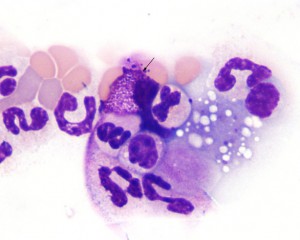
A well-granulated mast cell (arrow) is seen at the feathered edge of a blood smear from a dog with pancreatitis. The mast cell was attributed to the inflammation caused by the pancreatitis. The dog had no evidence of a mast cell tumor. When present in low numbers in blood, mast cells are usually carried out to the feathered edge (during smear preparation) but some can also be seen in the body of the smear during scanning at low power magnification (10x or 20x objective). Wright’s stain, 1000x magnification.

