
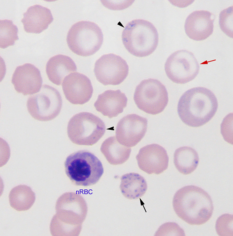
The anemia in the dog was moderate (29% hematocrit) and microcytic (64 fL) but not hypochromic (33 mg/dL). However, mild hypochromasia (increased central pallor due to a thin rim of hemoglobin) was evident in a subset of red blood cells in the smear (red arrow). The presence of siderocytes (blue dots in RBCs, arrowheads) are a clue that the hypochromasia may be due to inhibition of heme synthesis versus true or absolute iron deficiency. The dog also had a normoblastosis (139 nucleated RBCs [nRBC]/100 leukocytes) and marked regeneration (absolute reticulocyte count of 704,000/uL), which is excessive for the degree of anemia, another clue as to the presence of possible lead toxicity. The image shows an nRBC and basophilic stippling (black arrow on bottom of image; ribosome retention in red blood cells because lead poisoning inhibits the enzyme that breaks down ribosomes or pyrimidine 5′ nucleotidase) (modified Wright’s stain, 100x objective).