
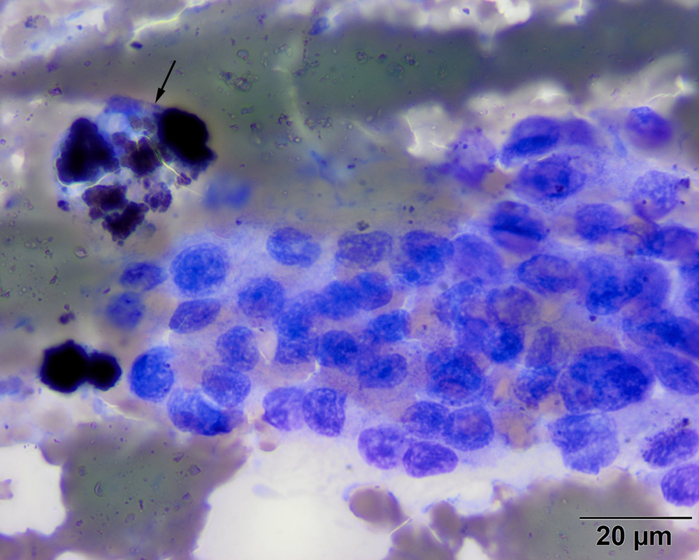
This image shows fine detail of the cells, illustrating their light to medium blue slightly grainy cytoplasm and nuclear and cellular uniformity. The latter features are characteristic of most endocrine or neuroendocrine tumors. Note the hemosiderin-laden macrophage, which also appears to have a phagocytized erythrocyte in the cytoplasm (arrow), indicating hemorrhage (modified Wright’s stain, 100x objective)