Aspirate of an esophageal nodule in a 1 year old dog
Case Information
A 1 year old neutered male Great Pyrenees mix was referred to Cornell University Hospital for Animal with a bicavitary (pleural and peritoneal) effusion that was diagnosed by the referring veterinarian. The dog had a three week history of inappetence after a dietary change. Clinical pathologic testing by the veterinarian revealed a panyhypoproteinemia (hypoalbuminemia and hypoglobulinemia). The dog did not respond to treatment with fenbendazole, metronidazole, and a probiotic. No other gastrointestinal signs, such as diarrhea or vomiting, were noted by the owners. On physical examination, the dog was tachycardic and tachypneic, with increased respiratory effort and abdominal breathing. A fluid wave was detected on abdominal palpation.
Serum chemistry testing confirmed a panhypoproteinemia (total protein concentration, 3.1 g/dL, reference interval [RI]: 5.5-7.2 g/dL; albumin concentration, 1.8 g/dL, RI: 3.2-4.1 g/dL; globulin concentration, 1.3 g/dL, RI: 1.9-3.7 g/dL). There was a concurrent hypocholesterolemia (116 mg/dL, RI: 136-392 mg/dL), mild hypomagnesemia (1.4 mg/dL, RI: 1.5-2.1 mg/dL), mild hyperglycemia (118 mg/dL, RI: 68-104 mg/dL), mild hypoferremia (79 ug/dL, RI: 97-263 ug/dL) and low total iron binding capacity (146 ug/dL, RI: 280-489 ug/dL) with a normal iron saturation (54%, RI: 27-66%). No relevant abnormalities were detected on a hemogram.
Thoracic and cranial abdominal radiographic imaging revealed mild bicavitary effusion. On abdominal ultrasonographic imaging, a peritoneal effusion and mildly hyperechoic intestinal mucosa were detected. An abdominocentesis yielded 3 ml of light yellow slightly cloudy fluid. The fluid had a total protein by refractometer of 1 g/dL and a nucleated cell count of 1.3 thou/uL. A differential cell count on cytospin smears of the peritoneal fluid consisted of 65% eosinophils, 21% small lymphocytes, 10% non-degenerate neutrophils, and 5% macrophages. A few mast cells were seen on scanning. Macrophages were leukophagocytic (neutrophils and eosinophils). Thoracocentesis yielded light yellow clear fluid with a total protein by refractometer of 0.5 g/dL and a nucleated cell count of 1.0 thou/uL. A differential cell count on cytospin smears of the pleural fluid consisted of 37% small lymphocytes, 25% segmented non-degenerate neutrophils, 23% macrophages, 14% eosinophils, and 1% mast cells. There were low numbers of mesothelial cells. Macrophages were leukophagocytic and erythrophagocytic.

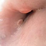
Endoscopic examination of the dog’s upper gastrointestinal tract revealed circular smooth nodules within the mucosa in the mid-esophagus at the level of the heart (Figure 1, see video).
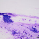
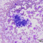
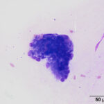
The stomach was normal in appearance but the duodenum had an abnormal granular appearance. The nodules in the esophagus were aspirated via endoscopic guidance and smears of the aspirates were submitted for cytologic evaluation (Figures 2-4). Endoscopic biopsies of the stomach and duodenum were taken for histopathologic examination. View the provided images then answer the questions below.
- Interpret the peritoneal and pleural fluid results (classify the effusions).
- What differential diagnoses would you consider as a cause for the effusions in this dog?
- What are the cells in the smears from the aspirates of the esophageal nodules and what is their likely tissue of origin?
- Do the esophageal nodules explain the effusions in this dog?
 |
 |
 |
Answers on next page
