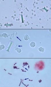
Upper panel: Low magnification view showing increased numbers of leukocytes and several struvite crystals (unstained wet prep). The leukocytes provide clear evidence of an inflammatory process; the background appears ‘busy’, but bacteria are not reliably identifiable at this magnification.
Middle panel: High magnification view of unstained wet prep showing leukocytes and clumps and chains of bacteria (arrows). Amorphous crystals or debris, however, can have a virtually identical appearance. Use of phase contrast microscopy can help in distinguishing between the two, but examination of a gram-stained drop of the urine sediment is most reliable.
Lower panel: High magnification view of a gram-stained slide. A neutrophil and gram positive cocci arranged in clusters and short chains are shown (arrows: some organisms have partially or completely decolorized). It can be concluded that the inflammatory process is caused or complicated by bacterial infection.

