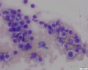
Figure 2: The round cells have moderate amounts of light gray/blue cytoplasm with low to moderate numbers of clear, discrete vacuoles. The nuclei are round to oval, central to eccentrically placed with finely stippled chromatin and indistinct nucleoli. Low numbers of inflammatory cells, including plasma cells (teal arrow) and eosinophils, were observed intercalated with the round cells. Wright’s stain, 50x.

