
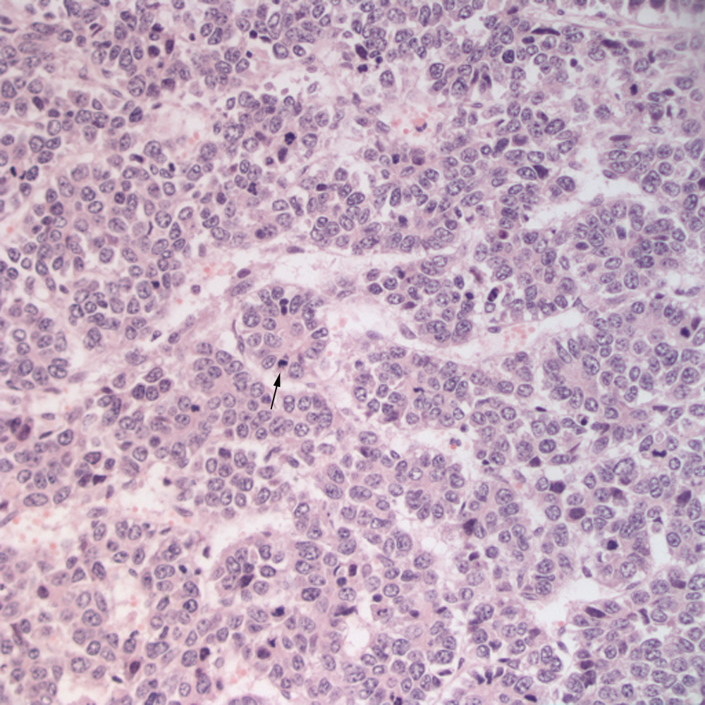
The tumor was composed of nests, lobules, and tubular- and acinar-like (arrow) arrangements. Note the mitotic figure at the tip of the arrow in the acinar-like arrangement. They were small to medium cells with paracentral nuclei and a moderate amount of eosinophilic cytoplasm. The mitotic rate was high (20x, H&E). This pattern fits with the histologic classification of the embryonal epithelial variant of hepatoblastoma.