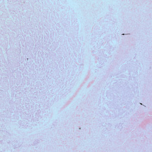
The liver contained multilobular masses of tumor cells, forming nests, lobules, and tubular-like and acinar-like arrangements (T). The masses were compressing the adjacent hepatic parenchyma (H), which was congested. Tumor masses were observed within veins in the portal regions (arrows) (5x objective, Hematoxylin & Eosin).

