
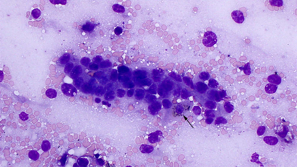
Scraping of a biopsy of a liver mass in a horse: Note the tubular arrangement of the tumor cells and the moderate anisokaryosis and mild to moderate anisocytosis. A single hepatocyte with green-brown pigment (bilirubin or lipofuscin presumptive) is closely intercalated with the tumor cells (arrow). Ruptured cells are in the background with one intact small lymphocyte (50x objective, Wright’s stain).