
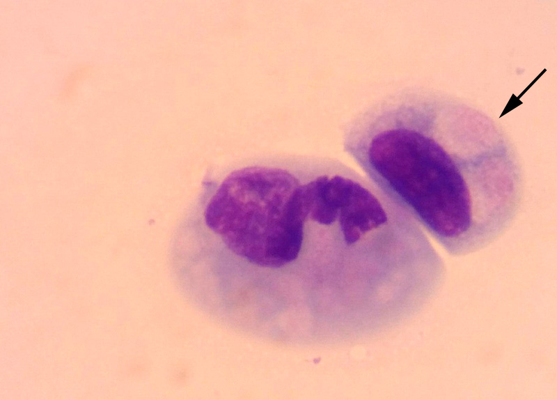
The two depicted cells are squamous epithelial cells from the conjunctival mucosa. There are two large pale diffuse inclusions with slight pink granularity within one of the epithelial cells (arrow). The adjacent cell may also contain inclusions (lower right of nucleus), but they are less distinct. The nucleus within the latter epithelial cell could be from a ruptured cell (Diff-quik).