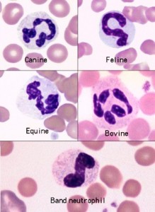
Canine neutrophils have white cytoplasm that contains small pink specific or secondary granules. Feline neutrophils have cytoplasm that is white and lacks visible granules. They may contain 1-3 small rod or round blue granules in their cytoplasm. These are Döhle bodies and are actually whorls of rough endoplasmic reticulum on electron microscopy. Equine neutrophils have white or slightly pink cytoplasm with no visible granules. The nuclei of equine neutrophils typically are long, thin and “knobby” with clumps of condensed chromatin projecting from the sides; they may not have classic segmentation. Ruminant neutrophils have white cytoplasm with small pink granules; these impart an overall pink tint compared to the other species. A band neutrophil from a dog is also shown. Compared to the segmented neutrophil, the band neutrophil has a uniformly smooth nuclear outline with no indentation (a segment is defined as a >50% constriction in the width of the nucleus). This band neutrophil is also not demonstrating toxic change.

