RBC analysis
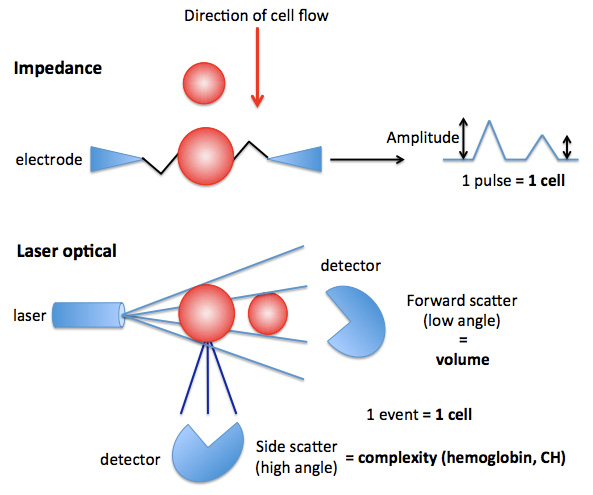
…ee of change in amplitude is a measure of the size (volume) of the RBC with smaller RBC causing less resistance and a lower amplitude. Laser-based hematologic analyzers allow RBC to pass through in single file through a flow cell containing a laser. As the RBC passes through the laser, it is counted as an event (providing a RBC count) but it also scatters the laser light. Two detectors can measure the scattered light. The first is the forward sca…