
Fine needle aspirate from a liver mass in a cat
Case information
A 10-year-old male castrated domestic shorthair cat initially presented to the referring veterinarian for fever, inappetence and an episode of vomiting. The cat was previously healthy and not on any medications. CBC, clinical chemistry profile and feline pancreatic lipase immunoassay (fPLI) were initially normal. The cat responded minimally to symptomatic treatment, including antibiotics, maropitant, NSAIDs and IV fluids. Over the course of several days, the cat decompensated further, becoming painful in the abdomen. Abdominal radiographs showed no significant findings and the cat was referred to Cornell University Hospital for Animals (CUHA) for further work up.
Upon initial exam at CUHA, the cat was persistently febrile at 103.2 F with a mildly increased respiratory rate and effort. A CBC showed a mild normocytic, normochromic, nonregenerative anemia of 26%. Although the overall leukocyte count (14.7 thou/uL, reference interval 5.1-16.2 thou/L) and neutrophil count were normal (9.4 thou/uL, reference interval 2.3-11.6), a mild left shift (1.2 thou/uL, reference interval 0-0.1 thou/uL), mild monocytosis (1.5 thou/uL, reference interval 0-0.7 thou/uL) and mild basophilia (0.3 thou/uL, reference interval 0-0.2 thou/uL) were evident. Clinical chemistry abnormalities included moderate hyponatremia and hypochloremia, moderate hypocalcemia, mild hypermagnesemia, moderate hypoalbuminemia and mild direct hyperbilirubinemia. Urinalysis showed marked proteinuria, mild glucosuria and marked bilirubinuria.
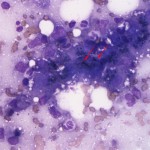
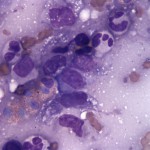
Overnight, the cat developed a further increased respiratory rate and respiratory effort. Thoracic imaging revealed a small amount of pleural effusion and 35 ml of fluid were removed from the right thorax. The fluid was analyzed and identified as a transudate (refractometer total solids <2.5 g/dL, nucleated cell count 0.4 thou/uL). An echocardiogram showed no cardiac abnormalities. Mild, diffuse hepatomegaly with ill-defined margins was noted on abdominal ultrasound, along with a mild peritoneal effusion. Within the liver, an approximately 1.2 x 2.6 x 2.8 cm mass was identified. Fine needle aspiration was performed of the liver mass and submitted for cytological evaluation. After examining the provided im
ages, try your hand at the following questions:
- What inflammatory components are present?
- Identify the black, linear structures indicated by the red arrows in Figure 2.
- Can an underlying cause for the inflammation be identified?
|
|
|
|

Answer on next page